Cervical Spine Anatomy: Facet Joint Zygoapophysial and Physiotherapy Graphics
Detailed visuals illustrating the anatomy of the cervical facet joints and their movements during flexion, extension, and lateral motions, with a focus on physiotherapy applications.
MedilawTV
204 views • Jul 18, 2011
About this video
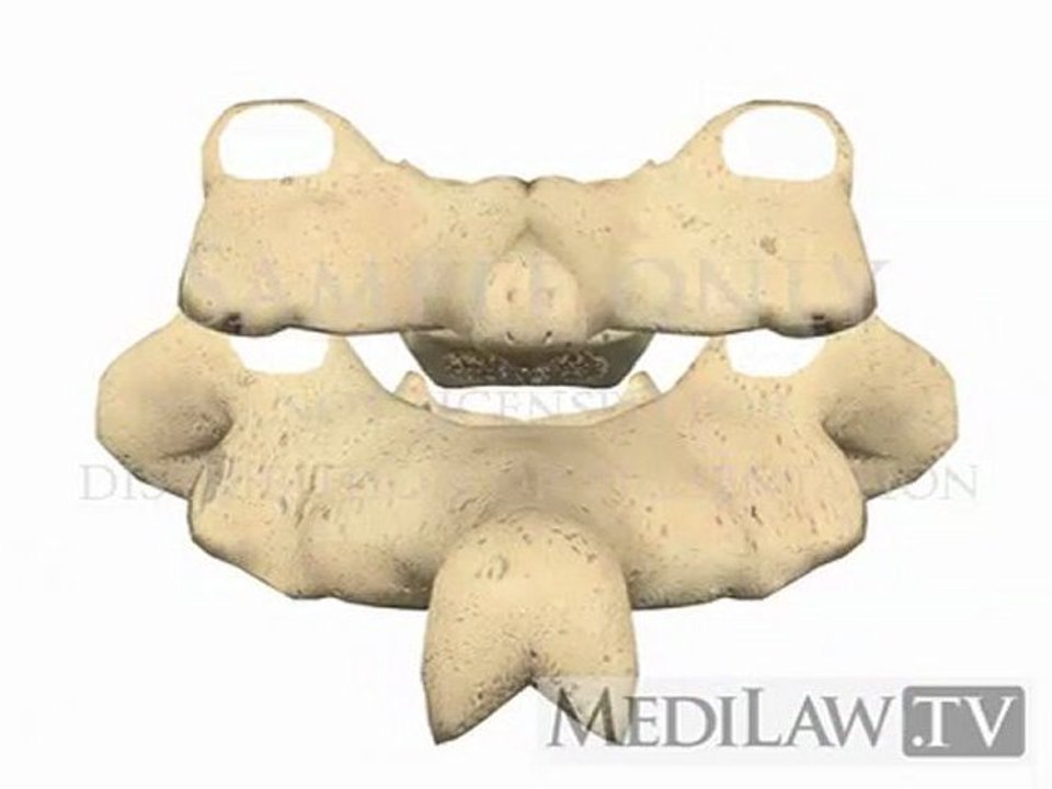
Cervical Spine Anatomy Facet Joint Zygoapophysial physiotherapy graphics. Shows the movements of the cervical facet joints during flexion-extension and lateral flexion. Also shows the medial branch nerves to the facet joints. An inferior articular process from the vertebra above moves against the superior articular process of the vertebra below to form a facet joint. This is also known as a zygo-apophysial joint. The bone ends on the joint are covered with a smooth cartilage, called hyaline cartilage. The facet joint is surrounded by a tough fibrous capsule. The inner lining of the capsule consists of the synovial membrane. The synovial membrane secretes and reabsorbs a small amount of synovial fluid. The synovial fluid lubricates and nourishes the joint surfaces. The adjacent vertebra are connected by the intervertebral disc in front of the spinal canal. Cervical Spine Anatomy Facet Joint Zygoapophysial physiotherapy graphics. Visit http://www.medilaw.tv for more information.
Video Information
Views
204
Duration
0:53
Published
Jul 18, 2011
Related Trending Topics
LIVE TRENDSRelated trending topics. Click any trend to explore more videos.
No specific trending topics match this video yet.
Explore All Trends