Detecting Squamous Cell Carcinoma with Advanced Soft X-Ray Microscopy 🔬
Discover how cutting-edge soft X-ray microscopy is revolutionizing the detection of squamous cell carcinoma, enabling detailed observation of biological samples in their natural state for early and accurate diagnosis.
Shpinetechnologies
13 views • Nov 29, 2014
About this video
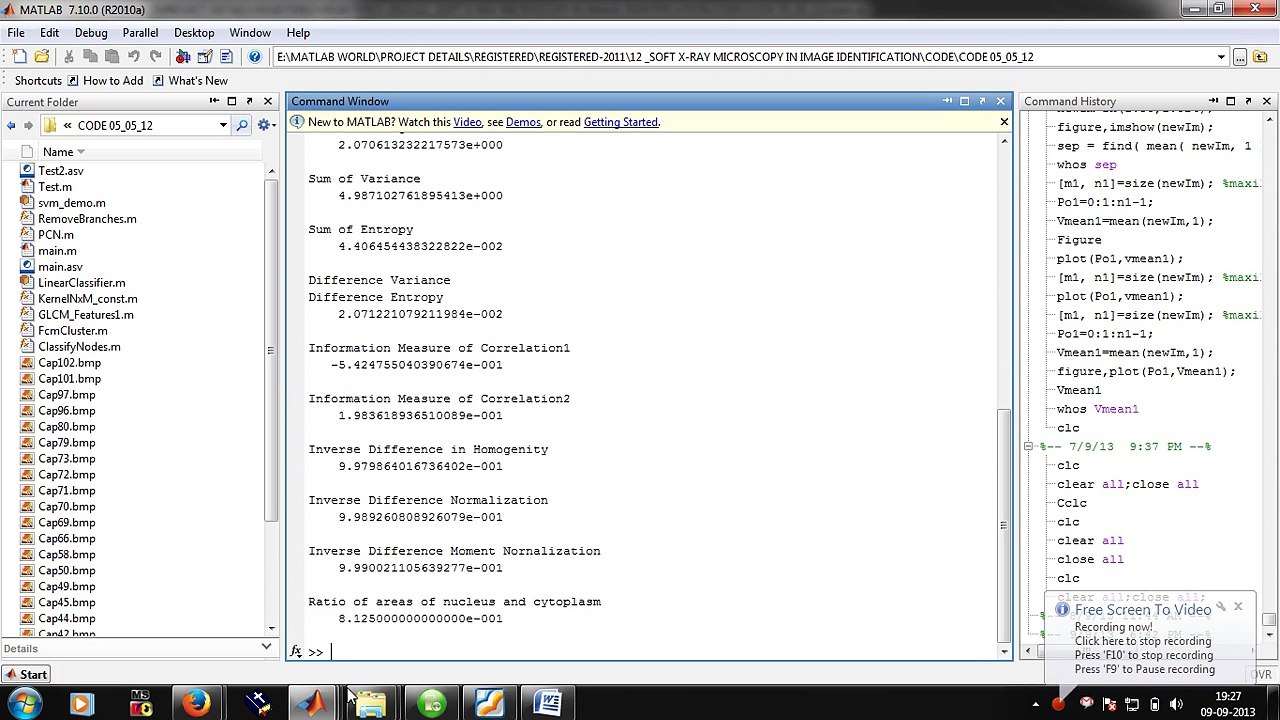
Nowadays, soft X-Ray microscopy technology is one of <br />the hottest methods that researches and observes the biological <br />samples of natural state, whose resolution is higher than the <br />imaging drawing of optical microscope and near to the imaging <br />drawing of electron microscope. The technology is simple for the <br />preparation of biological sample, which needs only <br />ultramicrocut. And the working procedure of traditional <br />pathology check, such as freezing, sealing wax, dehydration and <br />dyeing, etc. isn’t needed. A kind of image recognition system is <br />selected in the paper, which identifies and researches the tissue <br />morphological characteristics of ultrastructure imaging drawing <br />of squamocellular cancer of esophagus obtained by soft X-Ray <br />microscopy. The system mainly consists of components of image <br />preprocessing, image segmentation, cellular feature extraction <br />and cellular feature recognition, etc. Image preprocessing <br />includes grayscale transformation, histogram adjustment, etc. <br />Image segmentation includes segmentation based on the <br />threshold, image processing based on the structure morphology <br />of cell tissue, and edge detection, etc. In cellular feature <br />extraction, the method of extracting area connected is used. And <br />in cellular feature recognition, the judgment of area ratio of <br />nucleus and cytoplasm is adopted. According to the morphology <br />characteristics of tissue ultrastructure of squamocellular cancer <br />of esophagus, a kind of cancer identification method based on the <br />area threshold is employed emphatically in the paper, aiming at <br />distinguishing the normal cell and cancer cell successfully. The <br />technology application on the aspects of clinical diagnosis and <br />differentiate diagnosis of the image recognition system in the <br />paper still needs to be researched further
Video Information
Views
13
Duration
0:14
Published
Nov 29, 2014
Related Trending Topics
LIVE TRENDSRelated trending topics. Click any trend to explore more videos.
No specific trending topics match this video yet.
Explore All Trends